
Subacute stent thrombosis — the event occurring between 24 hours and 30 days after implantation — is the ideal teaching case for a cath lab team, because it forces a trainee to reason across pharmacology, procedural technique and human factors in one continuous decision chain. This subacute stent thrombosis module is built for a 60-minute facilitated session with fellows, cath lab nurses and technologists. It works equally well as self-directed study.
Module contents
Learning objectives for this subacute stent thrombosis module
By the end of this module the learner will be able to:
- Classify subacute stent thrombosis by timing and by ARC certainty category.
- Distinguish a pharmacological cause from a mechanical cause at the bedside, before angiography.
- Select appropriate loading and intraprocedural antithrombotic therapy for an unstable patient.
- State the intravascular imaging targets used to confirm adequate stent optimisation.
- Construct a discharge plan that reduces the risk of a recurrent event.
Case part 1: the call from the ward
A 58-year-old man underwent PCI to the proximal LAD eleven days ago — a 3.0 × 38 mm everolimus-eluting stent for a heavily calcified lesion. He was discharged on aspirin and clopidogrel. He now has crushing chest pain, is diaphoretic, and his blood pressure is 88/56.
Pause the session here. Ask the group three questions before revealing anything further.
- What is your working diagnosis and how confident are you?
- What do you want done in the next five minutes, in order?
- What single question would you ask the patient?
Teaching point
The single most informative question is when he last took clopidogrel. He stopped it four days ago because a dentist advised it before an extraction. Median time to subacute stent thrombosis after cessation within the first six months is about nine days, and this presentation sits squarely inside that window.
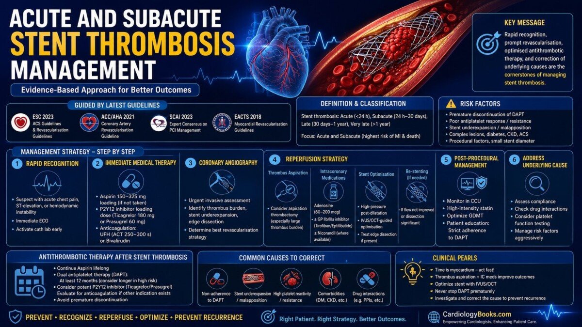
The second teaching point is about the loading choice. He thrombosed while taking clopidogrel; re-loading clopidogrel repeats an experiment that has already failed. Load aspirin 150–325 mg and ticagrelor 180 mg or prasugrel 60 mg, and anticoagulate with unfractionated heparin to an ACT of 250–300 seconds.
Case part 2: the angiogram
The proximal LAD is occluded at the stent with TIMI 0 flow and a large thrombus burden. The wire crosses easily.
Pause again. Ask what the group wants to do first, and what they specifically do not want to do.
Teaching point
Restore flow before imaging. An occluded segment gives an uninterpretable pullback and wastes contrast in a patient with borderline haemodynamics. Gentle balloon dilatation is usually sufficient.
Aspiration thrombectomy is reasonable here given a large, angiographically evident thrombus burden, but the group should be able to articulate why it is not routine: it reduces distal embolisation without reducing major adverse events, and routine use in randomised data carried a stroke signal. This is an important habit to teach — being able to defend a choice rather than perform it reflexively.
If oral absorption is unreliable in a vomiting or intubated patient, intravenous cangrelor gives immediate P2Y12 blockade with offset inside an hour. A glycoprotein IIb/IIIa inhibitor is an alternative bailout with heavy thrombus.
Case part 3: the imaging run
Flow is restored to TIMI 3. OCT pullback shows a minimum stent area of 3.6 mm² across a calcified segment with a calcium arc of 210° and thickness of 0.7 mm. Struts are apposed. There is no edge dissection and no neointimal coverage abnormality.
Pause. Ask the group to name the mechanism and the treatment, and to justify why re-stenting is not the answer.
Teaching point
This is underexpansion in a calcified lesion — the dominant mechanical cause, carrying roughly a thirteen-fold relative risk increase and accounting for about 26% of early events. The calcium features present here are exactly the triad that predicts underexpansion: arc ≥180°, thickness ≥0.5 mm and length over 5 mm.
The treatment is lesion modification and high-pressure post-dilatation with a non-compliant balloon, aiming for a minimum stent area of at least 4.5 mm² by OCT or 5.0 mm² by IVUS. Another stent layer adds strut burden without addressing the calcium that caused the problem.
Note for the group that this patient had two causes operating together — a missed mechanical problem at the index procedure and a pharmacological gap on day seven. Vignettes usually have one; real patients often have both.
Case part 4: closing the loop
Post-dilatation achieves a minimum stent area of 5.4 mm². Flow is TIMI 3 and the patient stabilises.
The discharge conversation is the part trainees skip, and it is the part that prevents the next admission. Aspirin continues indefinitely. Dual antiplatelet therapy runs at least 12 months, with a potent P2Y12 inhibitor rather than clopidogrel, and extension considered because a prior subacute stent thrombosis is itself a strong predictor of recurrence — roughly one in eight patients has a second one.
Ask the group who else needs to be told. The correct answer includes the dentist. Most interruptions come from another clinician acting on incomplete information, not from a non-adherent patient.
The subacute stent thrombosis algorithm
- Recognise. Chest pain within 30 days of PCI, ECG immediately, activate the lab. Do not wait for troponin.
- Load. Aspirin 150–325 mg plus ticagrelor 180 mg or prasugrel 60 mg. Heparin to ACT 250–300 s, or bivalirudin.
- Reperfuse. Wire, gentle balloon, selective aspiration for large thrombus burden. Intracoronary adenosine 60–200 mcg for no-reflow.
- Image. OCT or IVUS once flow is restored, to separate mechanical from pharmacological causes.
- Optimise. High-pressure post-dilatation to target minimum stent area. Cover dissection. Re-stent only when there is a genuine indication.
- Prevent. Potent P2Y12 inhibitor, ≥12 months of DAPT, high-intensity statin, risk-factor control, and an explicit interruption plan given to the patient and the referring physician.
Cath lab team skills checklist for subacute stent thrombosis
Use this as a direct observation tool during simulation or a real case debrief.
- Lab activated without waiting for biomarkers
- Correct loading agent chosen and rationale stated aloud
- ACT checked and documented before intervention
- Flow restored before imaging pullback
- Imaging findings verbalised to the team, not read silently
- Minimum stent area target stated and achieved
- Mechanism named explicitly in the procedure record
- Discharge antithrombotic plan written before the patient leaves the lab
- Interruption instructions communicated to patient and referrer
Facilitator notes
Run the case in four reveals rather than presenting it whole. The learning sits in the pauses, and the commonest facilitation error is showing the OCT image before the group has committed to a mechanism.
Expect two predictable wrong answers. The first is re-loading clopidogrel — challenge it by asking what evidence exists that this patient inhibits platelets adequately on clopidogrel. The second is implanting a second stent — challenge it by asking what the new stent does to the calcium.
Allow ten minutes at the end for the human-factors discussion. Ask each participant to describe how their own unit communicates antiplatelet interruption to dentists, endoscopists and surgeons. This usually surfaces a real system gap, and it is the highest-value ten minutes of the session.
Assessment questions
- Define subacute stent thrombosis by timing and mechanism, and state the ARC certainty category for an unexplained death at day 25 without angiography.
- A patient presents on day 12 having stopped his P2Y12 inhibitor on day 5. Name the dominant mechanism and the loading regimen you would use.
- State the minimum stent area targets for OCT and IVUS.
- List the three OCT calcium features that predict underexpansion.
- Give two reasons not to implant a second stent when imaging shows isolated underexpansion.
- Name two trials supporting imaging-guided PCI and state what each showed.
- Outline the discharge antithrombotic plan and name the one instruction the patient must remember.
Model answers are provided in the facilitator pack for enrolled course participants.
Related: Browse all cardiology courses · Cardiology Learning Center · Mentorship programme
References. Stent Thrombosis: A Contemporary Guide to Definitions, Risk Factors, and Management. Front Cardiovasc Med 2025;12:1622235. · 2025 ACC/AHA/ACEP/NAEMSP/SCAI ACS Guideline, Circulation 2025. · RENOVATE-COMPLEX-PCI · ADAPT-DES · TOTAL trial core-lab analysis, EuroIntervention.